Hi, what do you want to do?
Exploratorium
Exploratorium: Microscope Imaging Station: Mouse Embryonic Stem Cells
Stem cells have an enormous impact on the structure of a living creature. This site features still images as well as short videos of these cells in action.
Museum of Science
Mo S: Image Gallery of the Scanning Electron Microscope
At this resource you'll find everything from a mosquito head to a fly foot, from a dentist's drill to toilet paper! These and just about everything in between as seen through a scanning electron microscope.
CK-12 Foundation
Ck 12: Plix: Microscopes
[Free Registration/Login Required] Focus a virtual microscope and learn about the images microscopes produce on this site. Also included is a short quiz on microscopes.
TeachEngineering
Teach Engineering: Imaging Dna Structure
Learners are introduced to the latest imaging methods used to visualize molecular structures and the method of electrophoresis that is used to identify and compare genetic code (DNA). Students should already have basic knowledge of...
Exploratorium
Exploratorium: Microscope Imaging Station: Cell Development
This interesting site provides videos and photos of cells going through the process of cell division. Observe Zebrafish development and Sea urchin embryo cell division in video format.
Exploratorium
Exploratorium: Microscope Imaging Station: Cell Motility
This interesting site explains how cells move and provides videos depicting the movement of various human and animal cells.
Exploratorium
Exploratorium: Microscope Imaging Station: Blood Cells
This colorful site provides a gallery of blood cell photos. You can observe human white blood cells, human red blood cells, sheep red blood cells, and zebrafish red blood cells. As an added bonus, you can enlarge the red blood cell...
Florida State University
Florida State University: Magnification Module
Explore the effect of increasing the magnification on a microscope when viewing various samples such as onion root mitosis.
Other
Scimat: Foods Under the Microscope
A technical site that describes different types of microscopes and delves into the chemical makeup of milk, yogurt, various various types of microorganisms. Impressive pictures supplement the text. Links to images of microorganisms.
Florida State University
Florida State University: Molecular Expressions: Basic Concepts in Optical Microscopy
This site explores the microscope in great detail. Content includes a focus on the microscope's basic functions (including a general diagram), the history of the microscope, each feature of the microscope, differing objectives,...
Arizona State University
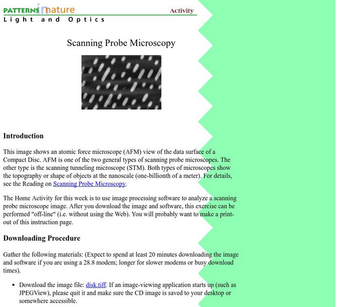
Arizona State University/spm Home Activity
Investigate a SPM image at home with this activity of the week. Links back to more info about SPM's.
Other
University of Muenchen: Scanning Probe Microscopy Group
Scroll down to research to find images of scanning tunneling and atomic force microscopes.
Other
Nikon: Nikon Small World: Photomicrography Competition
Nikon has, for years, sponsored a competition to showcase the work of photographers who take pictures of microscopic subjects. View galleries of winning images for past competitions as you learn about the art and science of microscopy.
Curated OER
Science Kids: Science Images: Lung Fibers
A highly detailed electron microscope image of collagen fibers found in the lung.
Curated OER
Using a Compound Microscope (Photo by Paul Billiet)
This site is a step by step instructions on how to use a microscope. Begins with the proper way to get the microscope and ends with putting the microscope. The correct way to look at slides and care for you microscope are also discussed....
Curated OER
Compound Microscope Showing the Stage and Spring Clips
This site is a step by step instructions on how to use a microscope. Begins with the proper way to get the microscope and ends with putting the microscope. The correct way to look at slides and care for you microscope are also discussed....
Curated OER
A Compound Microscope
Learn how to calculate the total magnification of a microscope on this concise site. Links to making a slide, questions on the use of the microscope, and other related microscope topics are included on this site.
University of Kansas Medical Center
University of Kansas Medical Center: Cell Structure
Discover microscopic images of various eukaryotic cells, both plant and animal.
University of Kansas Medical Center
University of Kansas Medical Center: Bone
See several specimens of microscopic images of bone cells and tissue.
University of Kansas Medical Center
University of Kansas Medical Center: Basic Histopathology
These microscopic images of different cells and tissues from the human organs gives an idea of some the pathological processes that occur and the importance of histology.
Lawrence Berkeley National Laboratory
Berkeley Lab: Ncem: Electron Micrographs
This site contains a gallery of scanning electron micrographs of non living substance.
Iowa State University
Iowa State University: Scanning Electron Microscopy
Information on the scanning electron microscope and how it works, accompanied by a library of images.
Other
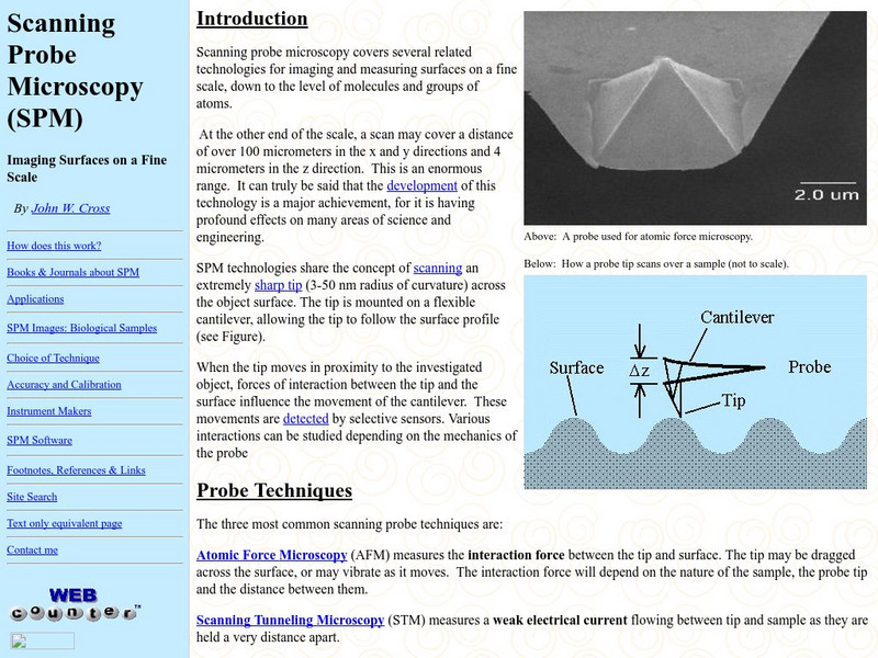
Scanning Probe Microscopy
This site provides a fairly in depth description of Scanning Probe Microscopes as well as links and images. This site is designed for those with a strong understanding of this field.


