Hi, what do you want to do?
University of Minnesota
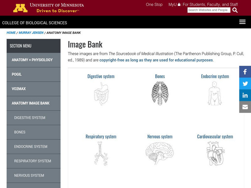
University of Minnesota: Anatomy Image Bank
Hundreds of anatomy diagrams for classroom or medical use. Each image represented is a simple black-and-white professional drawing of a specific part of the human body.
Other
Zygote: Zygote Anatomy Collection
A 3D anatomical product that allows students to explore the muscles, bones, organs, skin, and blood vessels that compose the human body.
eSkeletons
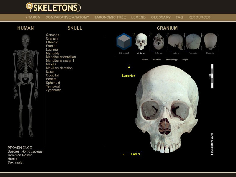
E Skeletons: Human
Studying the skeletal parts of a human? Click on various parts of the human skeleton and proceed to select the items to view in detail.
Cosmo Learning
Cosmo Learning: Video: 3 D View of Human Female Anatomy
A video animation with music exploring the workings of the female body. This 3D views allows you to get a glimpse of muscles, organs, and bone structure of the female body. Video shows you the network of veins and arteries of the body as...
Cosmo Learning
Cosmo Learning: Video: 3 D View of Human Male Anatomy
A video animation with music exploring the workings of the female body. This 3D views allows you to get a glimpse of muscles, organs, and bone structure of the male body. Video shows you the network of veins and arteries of the body as...
National Institutes of Health
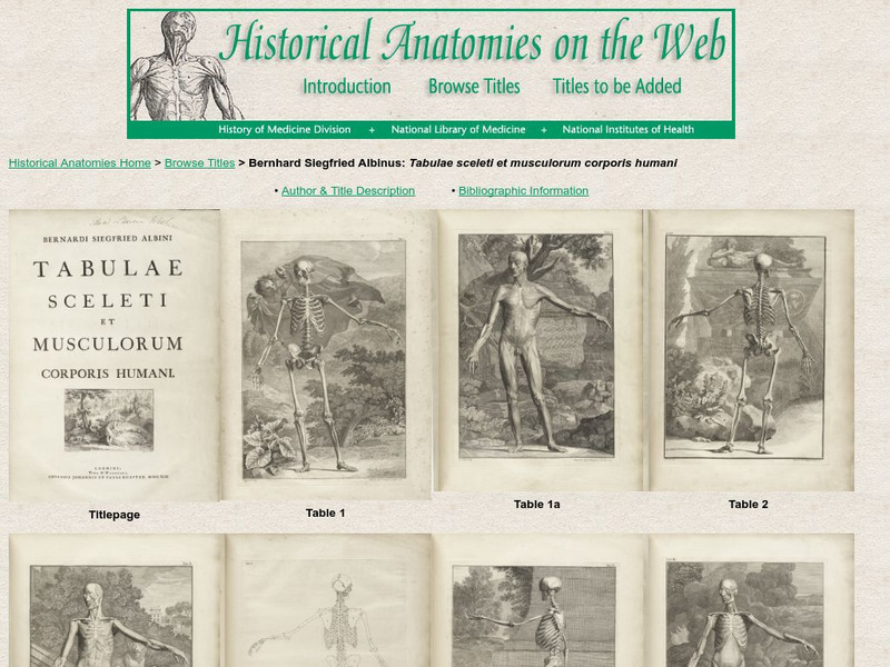
National Library of Medicine: Historical Anatomies of the Web: Albinus, Bernhard
Images from the 18th century anatomy text, "Tabulae sceleti et musculorum corporis humani" by Bernhard Siegfried Albinus. There are drawings of individual bones, skeletons and musculature. The text on the images is in Latin. Title and...
Biology Corner
Biology Corner: Anatomy: Skeletal System: Flash Cards
Printable collection of drawings of bones in the human body, suitable for testing knowledge of particular structures.
National Cancer Institute at the National Institutes of Health
Seer Training Modules: Introduction to the Skeletal System
Self-guided learning activity where students learn about the structure and function of the human skeletal system. There is a short quiz at the end of the lesson to check for understanding.
ClassFlow
Class Flow: Anatomy, Appendicular Skeleton
[Free Registration/Login Required] This flipchart presents the names of the bones in the human body. It includes an activity in which students label the bones after they have studied them.
Curated OER
Albinus Table 1 A
Images from the 18th century anatomy text, "Tabulae sceleti et musculorum corporis humani" by Bernhard Siegfried Albinus. There are drawings of individual bones, skeletons and musculature. The text on the images is in Latin. Title and...
Curated OER
Albinus Table 1 A
Images from the 18th century anatomy text, "Tabulae sceleti et musculorum corporis humani" by Bernhard Siegfried Albinus. There are drawings of individual bones, skeletons and musculature. The text on the images is in Latin. Title and...
Curated OER
Albinus Table 2 A
Images from the 18th century anatomy text, "Tabulae sceleti et musculorum corporis humani" by Bernhard Siegfried Albinus. There are drawings of individual bones, skeletons and musculature. The text on the images is in Latin. Title and...
Curated OER
Albinus Table 2 A
Images from the 18th century anatomy text, "Tabulae sceleti et musculorum corporis humani" by Bernhard Siegfried Albinus. There are drawings of individual bones, skeletons and musculature. The text on the images is in Latin. Title and...
Curated OER
Albinus Table 5
Images from the 18th century anatomy text, "Tabulae sceleti et musculorum corporis humani" by Bernhard Siegfried Albinus. There are drawings of individual bones, skeletons and musculature. The text on the images is in Latin. Title and...
Curated OER
Albinus Table 5
Images from the 18th century anatomy text, "Tabulae sceleti et musculorum corporis humani" by Bernhard Siegfried Albinus. There are drawings of individual bones, skeletons and musculature. The text on the images is in Latin. Title and...
Curated OER
Albinus Table 7
Images from the 18th century anatomy text, "Tabulae sceleti et musculorum corporis humani" by Bernhard Siegfried Albinus. There are drawings of individual bones, skeletons and musculature. The text on the images is in Latin. Title and...
Curated OER
Albinus Table 7
Images from the 18th century anatomy text, "Tabulae sceleti et musculorum corporis humani" by Bernhard Siegfried Albinus. There are drawings of individual bones, skeletons and musculature. The text on the images is in Latin. Title and...
Curated OER
Albinus Table 8
Images from the 18th century anatomy text, "Tabulae sceleti et musculorum corporis humani" by Bernhard Siegfried Albinus. There are drawings of individual bones, skeletons and musculature. The text on the images is in Latin. Title and...
Curated OER
Albinus Table 8
Images from the 18th century anatomy text, "Tabulae sceleti et musculorum corporis humani" by Bernhard Siegfried Albinus. There are drawings of individual bones, skeletons and musculature. The text on the images is in Latin. Title and...
Curated OER
Albinus Table 9
Images from the 18th century anatomy text, "Tabulae sceleti et musculorum corporis humani" by Bernhard Siegfried Albinus. There are drawings of individual bones, skeletons and musculature. The text on the images is in Latin. Title and...
Curated OER
Albinus Table 9
Images from the 18th century anatomy text, "Tabulae sceleti et musculorum corporis humani" by Bernhard Siegfried Albinus. There are drawings of individual bones, skeletons and musculature. The text on the images is in Latin. Title and...
